*** NOTE ***
The Journal online has become part of Concordia University NOW, your source for the latest university news and upcoming events. This site will no longer be updated. Visit the NOW website to read the Journal online and more.

September 11, 2008 | Vol. 4, No. 1
By Wendy Smith
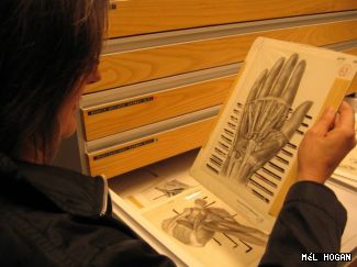
Kim Sawchuk cradles one of the seventy-odd year-old illustrations from the original Grants collection. Rendered in precise detail down to the last knuckle crease, an outstretched hand is drawn with the skin partially peeled back, exposing highways of veins swerving around muscle and bone. The image appears to have been lit from within, a quality achieved by applying layers of carbon dust to special film.
Decades before macabre images of disease and bodily trauma hit our television screens on shows like CSI and House, medical illustrators showed people what it would be like to travel inside their bodies. Now, four Concordians are setting out to study those pioneers who depicted the world beneath our skin.
Ask any physician about the tome he or she consulted most frequently during those years of training, and chances are two names will come up: Grays, the classic textbook on human physiology, and Grants, the first North American medical atlas that was originally published in 1943 and, now in its twelfth edition, continues to reign as the world standard.
The Illustrating Medicine project will initiate research on Grants Atlas specifically on the illustrators who made significant contributions to the field of medical education.
We know very little about these illustrators, said principal investigator Kim Sawchuk, associate professor in Concordias Department of Communication Studies. Atlases were named after the anatomist, not the illustrator. The images are central to any anatomical atlas, yet the illustrator often was eliminated from historical memory.
But traces of documentation about the illustrators still remain and Sawchuk and her team plan to hunt down these bits of information and integrate them with digital renderings of the original drawings in a searchable, online archive.
What we do know about the medical illustrators of the 1940s, says Sawchuk, is that they were unique. They practiced a discipline that straddles the divide between art and science, demanding proficiency with a paintbrush and a profound knowledge of anatomy. And at a time when men dominated other corners of medicine, the majority of illustrators were women.
Were bringing these women out of the shadows, said Nancy Marrelli, project participant and director of the Concordia Archives.
Illustrating Medicine, which recently received a three-year, $107,000 grant from the Social Sciences and Humanities Research Council, brings together an interdisciplinary team of scholars from Concordia and the University of Toronto. The Concordia contingent consists of Sawchuk; Marrelli; Mél Hogan, a candidate in the Joint Doctorate in Communication; and Nina Czegledy, associate adjunct professor in the Faculty of Fine Arts. They are joined by U of T researchers Nick Woolridge and Brian Sutherland.
Czegledys Digitized Bodies, Virtual Spectacles exhibition was the catalyst for this project. The first show to present the work of early Canadian medical illustrators as art in its own right, it toured Canada and Budapest in 2000. When Sawchuk marvelled over the anatomical drawings on display, Czegledy told her, Just wait until you see the rest of them! she recalled. And they are beautiful.
These illustrations are severely vulnerable to heat, light, dust, excessive handling even the oils in human skin are potentially destructive. Sawchuk didnt feel comfortable even handling them without consulting a professional archivist, because of the possibility of unintentional damage.
So she recruited Marrelli, who promptly paid a site visit to the bowels of the University of Toronto medical school, where the fragile drawings are languishing in inappropriate, non-archival storage cabinets, all jumbled up, with bits of tape stuck to them.
If you had told me Id be sitting in this museum surrounded by body parts, I would never have believed you, Marrelli confessed. But theres a certain curiosity about the whole thing. This is archival material, it has great historical value, and its not really recognized.
For me, this project is the missing link, agreed Sawchuk, whose previous research has investigated various aspects of feminism, biomedicine and embodiment. There have not been adequate histories of Canadian medical illustration. And medical history is part of our cultural history.
Next issue: September 25, 2008

Page Title: Anatomically correct research project - Concordia University - Montreal, Quebec, Canada
Page URL:http://cjournal.concordia.ca/archives/20080911/anatomically_correct_researc...oject.php
Date Printed: Mon July 27, 2026